
Research grants
Retina UK aims not only to progress research along established threads, but to stimulate new thinking, encourage innovative approaches and nurture original ideas.
Our Research Grants support projects of varying length which seek to explore new ideas or test new theories. We have made a commitment to support the talented research teams who are delivering the ground-breaking projects listed below. We are extremely grateful for each and every donation we receive.
Current grants
- Lead researcher: Professor Tim Curtis
- Location: Queen’s University of Belfast, Northern Ireland
- Type of funding: PhD studentship
- Total funding: £119,980
- Duration: January 2027 – January 2030
- Co-funded with the Macular Society
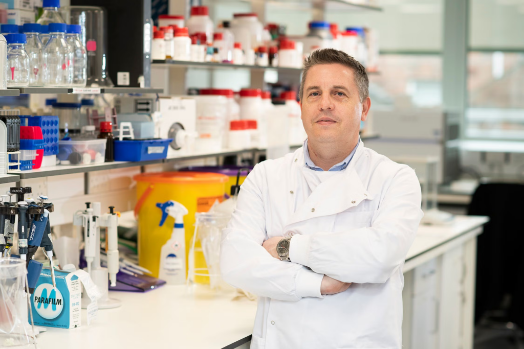
Professor Tim Curtis, lead researcher (QUB/PA).
The challenge
Inherited macular dystrophies are a group of diseases that affect the macula. This tiny area of the retina is only about the size of the rubber on the end of a pencil. But the macula gives us our central vision and lets us see in colour and fine detail. When a person’s macula breaks down, it becomes very difficult for them to read, use digital screens, or recognise faces. Inherited macular dystrophies include Stargardt disease, Best disease, and central areolar choroidal dystrophy. These conditions often begin during childhood or young adulthood, so they can seriously impact education, employment, and mental health.
There are currently no treatments available for inherited macular dystrophies. Because there are many different gene faults that can cause these conditions, researchers are searching for treatments that could work regardless of the genes involved.
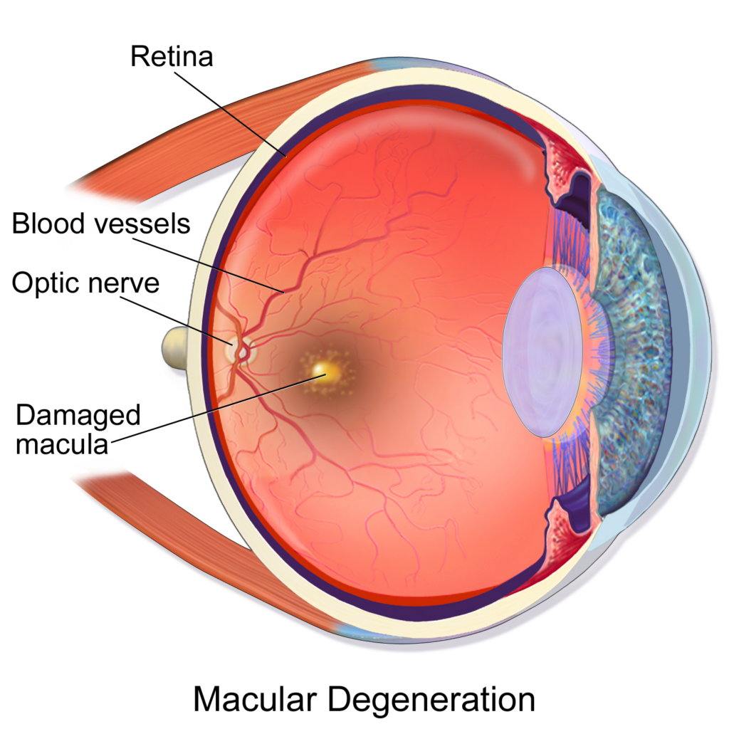
The position of the macula in the eye (BruceBlaus, CC BY-SA 4.0).
The vision
Although inherited macular dystrophies and diabetes are very different conditions, both can lead to the build-up of harmful molecules called aldehydes in the retina. These molecules are produced naturally by the body but can accumulate when the retina is under stress. Over time, these molecules can damage cells in the retina and contribute to vision loss.
Professor Tim Curtis’ team recently discovered that adding a small molecule called 2-HDP to drinking water can protect rats with diabetes from retina damage. The drug seems to work by hoovering up toxic aldehydes before they can do too much damage. Because similar aldehydes build up in inherited macular dystrophies, the researchers think 2-HDP could help slow the damage they cause in conditions such as Stargardt disease.
To test this theory, the researchers will first need to find out where and when toxic aldehydes build up as inherited macular dystrophies develop. They will carry out this work in mice bred to develop similar symptoms to these conditions. Once the team have drawn up this map of aldehydes in the macula, they will be able to see if treating the mice with 2-HDP changes it. They will also measure the levels of stress, inflammation, and damage in the macula, and test whether 2-HDP treatment can slow vision loss in these mice.
What could this mean for our sight loss community?
This laboratory research is still at an early stage and will not lead directly to a treatment. However, if successful, this work could pave the way for a new treatment approach to slow disease progression. Unlike gene therapies, which only tackle a single genetic cause, this approach could potentially benefit people with a variety of inherited macular dystrophies.
Progress so far
Check back in early 2028 for one-year updates on the project.
Learn more about this research
Read about Professor Tim Curtis’ other research
Read about the Macular Society, who are co-funding this project
Read about the most common types of inherited macular dystrophy
Read our guide to Stargardt disease
Read our guide to Best disease
Read our guide to central areolar choroidal dystrophy
Read our guide to X-linked retinoschisis
Read our guide to Doyne honeycomb dystrophy
Read our guide to Pattern dystrophy
Read about the impact inherited macular dystrophies can have on wider life
Read about the gene faults that can cause inherited macular dystrophies
Read about sight loss treatments that may work regardless of the genes involved
Read about Professor Tim Curtis’ work with 2-HDP in diabetic rats
- Lead researcher: Professor Luminita Paraoan
- Location: Manchester Metropolitan University, England
- Type of funding: Short innovation grant
- Total funding: £50,000
- Duration: January 2026 – April 2026

Professor Luminita Paraoan, lead researcher (Edge Hill University).
The challenge
As we get older, every part of our body changes, including the eye. Ageing cells in the retina become less efficient at generating energy, communicating with one another, and cleaning up toxic waste products. These changes make the retina more vulnerable to disease.
Many of these age-related changes are also seen in inherited retinal dystrophies, a group of conditions that cause progressive sight loss. In fact, for many people with inherited retinal dystrophies, their vision keeps getting worse with age. Yet surprisingly little research has explored how the ageing process affects inherited retinal diseases.
The vision
In Manchester, Professor Paraoan’s team is discovering whether ageing and inherited sight loss affect the same biological pathways. Targeting these shared pathways could provide entirely new ways of treating inherited retinal dystrophies.
The team will use a gene-editing technology called CRISPR (clustered regularly interspaced short palindromic repeats) on retina cells grown in the laboratory. By binding and cutting specific regions of DNA, this tool allows scientists to switch off genes one by one and observe the effects on retina cells. Professor Paraoan will carry out a large-scale screen, testing thousands of genes.
If you wanted to understand how an engine works, you could remove one component at a time and see what happens. When the engine failed, you’d know that the part you just removed was important. CRISPR screening works in a similar way. By switching off genes one by one, researchers can identify which ones help retinal cells stay healthy and which genes contribute to disease.
The team will perform these experiments on retina cells carrying faults in genes linked to several inherited retinal conditions, including Stargardt disease, Usher syndrome, Best disease, and Leber congenital amaurosis. They will then artificially “age” some of the cells and compare the results. This will allow the researchers to identify genes and biological pathways that influence disease, ageing, or both.
By finding genes that protect retinal cells from the combined effects of ageing and inherited disease, the research could uncover promising targets for future therapies. Equally, genes that make cells more vulnerable could highlight biological processes that should be blocked or modified.
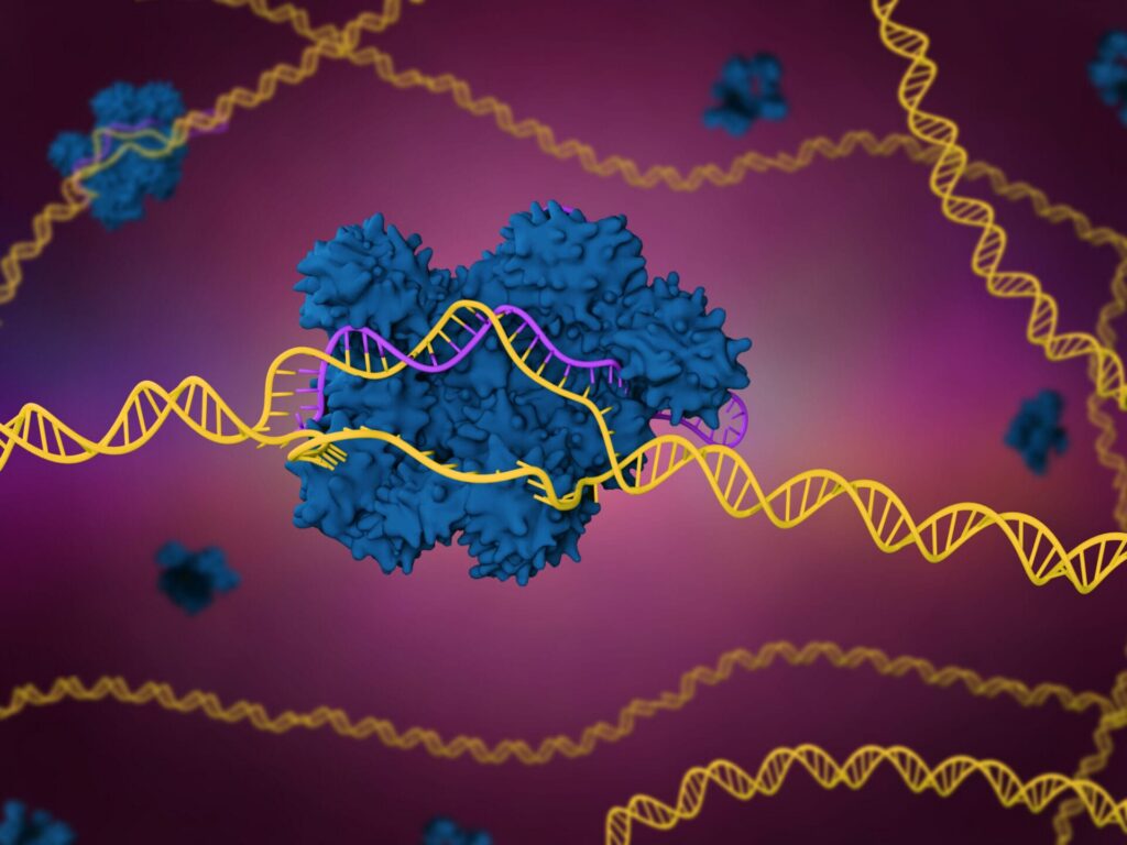
A computer-generated representation of a CRISPR protein (blue) bound to a specific gene (DNA shown in yellow). Professor Paraoan is using CRISPR to switch genes off in retina cells to find out which genes help keep the retina healthy during ageing. (Meletios Verras, Getty Images).
What could this mean for our sight loss community?
This project will not directly lead to a new treatment. However, it could reveal entirely new ways of thinking about inherited retinal dystrophies.
Importantly, any treatments developed from these findings could potentially work across multiple inherited retinal dystrophies, regardless of the specific gene fault responsible. This means they could benefit a much wider group of people than current gene-specific approaches.
The project will also provide some of the first detailed insights into how ageing affects inherited retinal diseases, an area of research that has received very little attention despite its importance to people living with progressive sight loss.
Progress so far
Check back in early 2027 for the final outcomes of this project.
Learn more about this research
Read more about Professor Luminita Paraoan’s research
Read our guide to Stargardt disease
Read our guide to Leber congenital amaurosis
Read our guide to Usher syndrome
Read our guide to Best disease
Read about the gene faults that can cause inherited retinal dystrophies
Read about sight loss treatments that may work regardless of the genes involved
- Lead researcher: Doctor Tess McCann
- Location: University of Dublin, Ireland
- Type of funding: Short innovation grant
- Total funding: £50,000
- Duration: October 2025 – February 2026
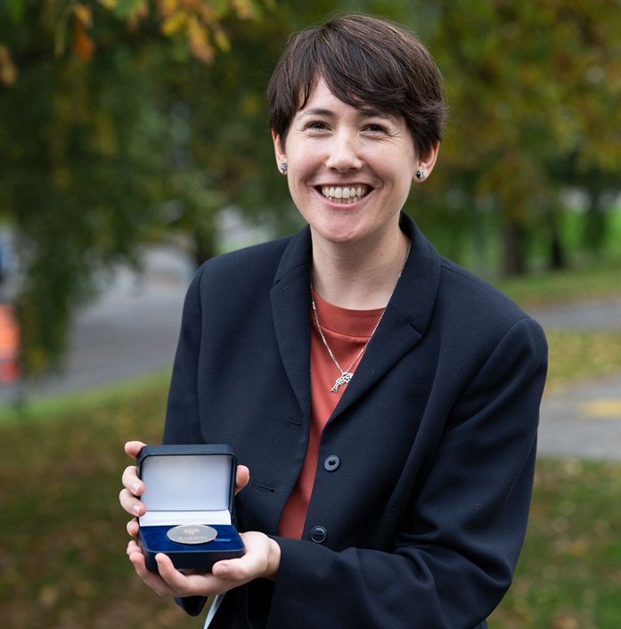
Doctor Tess McCann, lead researcher (UCD).
The challenge
Inherited retinal dystrophies are a group of diseases that affect the retina. This area, found at the back of the eye, is only about the size of a postage stamp. But the retina converts light into electric signals which are sent to the brain, allowing us to see. In many inherited retinal dystrophies, the light-sensing cells in a person’s retina die off over time, causing gradual vision loss. Once these cells are lost, they aren’t naturally replaced, which makes this vision loss irreversible.
There’s currently only one treatment available on the NHS for inherited retinal dystrophies, and it only applies to a very small proportion of people with these conditions. Because there are many different gene faults that can cause inherited retinal dystrophies, researchers are searching for treatments that could work regardless of the genes involved.
The vision
Doctor Tess McCann has spent her career studying a two-inch iridescent fish commonly found in pet shops around the world. The zebrafish, known as Danio rerio to scientists, has the remarkable ability to repair its retina. When zebrafish lose their light-sensing photoreceptor cells, another type of cell in the retina generates replacement photoreceptors to restore lost sight.
Good for the fish, but how is it relevant to us? In the last decade or so, scientists have learned that zebrafish are closer on our family tree than we thought. More than eight in every ten human genes linked to disease have counterparts in zebrafish. So, if we can understand how zebrafish manage to repair their retinas, could we kickstart the same process in people with inherited retinal diseases?
That’s what Doctor McCann is exploring in Dublin. Her team is taking detailed snapshots of the proteins found in injured zebrafish retinas as they repair themselves. Proteins are the molecules that do most of the work in cells. There are about 26,000 different proteins in zebrafish, with jobs like digesting food, moving muscles, and detecting light. By identifying which proteins are present and switched-on during retina repair, Doctor McCann hopes to reveal pathways that are also present in human eyes. If successful, this work could point the way to an entirely new approach for treating the damage in inherited retinal dystrophies.
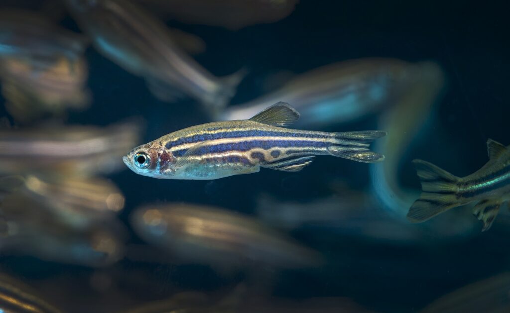
A zebrafish in a research laboratory (Stephen Ward, Oregon State University).
What could this mean for our sight loss community?
This laboratory research is still at an early stage and will not lead directly to a treatment. However, one of the biggest barriers to developing treatments that repair the retina is understanding why mammal retinas don’t do this naturally. By comparing proteins in zebrafish and mouse retinas, Doctor McCann’s team hopes to close that knowledge gap.
If successful, this research could set the stage for new treatment approaches to regenerate damaged retinas. Unlike gene therapies, which only tackle a single genetic cause, this strategy could potentially benefit people with a variety of inherited retinal dystrophies.
Although much more work is needed before any therapy could be developed, this project could provide important clues for restoring vision by stimulating the retina’s dormant repair mechanisms.
Progress so far
Check back at the end of 2026 for the final outcomes of this project.
Learn more about this research
Read more about inherited retinal dystrophies
Read about Doctor Tess McCann’s prize-winning research
Read more about how the zebrafish repairs its retina
Read about the similarities between human and zebrafish genes
Read about the 26,000 zebrafish proteins
Read about a clinical trial aiming to repair the retina with transplanted cells
Read about a biotech company aiming to unlock retina repair in another way
Read about other sight loss treatments that may work regardless of the genes involved
- Lead researcher: Professor Jane Sowden
- Location: University College London, England
- Type of funding: Project grant
- Total funding: £327,530
- Duration: December 2024 – November 2027
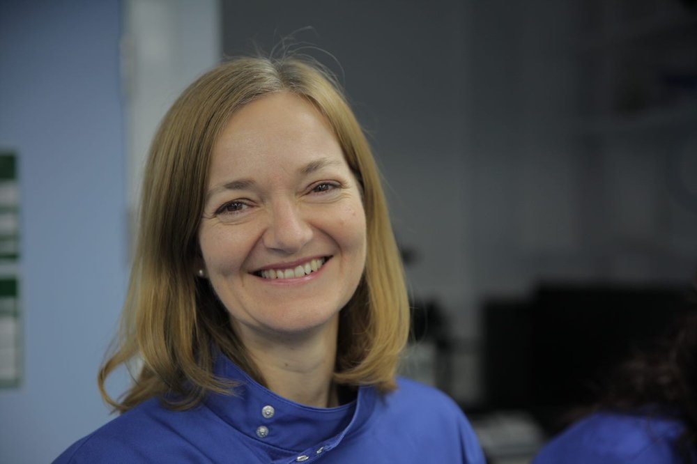
Professor Jane Sowden, lead researcher (Great Ormond Street Hospital).
The challenge
Faults in more than 300 genes are known to cause inherited retinal dystrophies. Researchers are developing gene therapies for some of these conditions, which aim to replace the fault gene with a healthy copy. However, most people with inherited sight loss still don’t have a treatment available for their gene fault. This is particularly true for people with advanced sight loss who have already lost most of their retina’s light-sensing cells.
To see, we rely on millions of light-sensing cells passing signals to the brain. Once these cells die, the retina cannot naturally replace them. One possible solution is to grow new light-sensing cells in the laboratory and transplant them into the eye. Scientists have already found ways of growing light-sensing cells in the laboratory from stem cells. Studies have shown that these light-sensing cells can survive after transplantation and restore some visual function in animas with retinal disease. Early-stage human trials of light-sensing cell transplants are also underway.
However, there are still important obstacles to overcome. Delivering cells to the retina is challenging. Many cells can be damaged during transplantation, fail to organise correctly, or struggle to survive in sufficient numbers after they’re delivered.
The vision
In London, Professor Sowden’s team is developing a tiny patch to help replacement cells reach the retina safely and survive after transplantation.
The researchers will first grow healthy light-sensing cells from stem cells in the laboratory. They’ll then place these cells onto a biodegradable scaffold: a microscopic framework that helps keep the cells in the correct arrangement. The finished patch, which will be about the size of a grain of rice, will contain thousands of replacement cells.
Imagine trying to grow a crop of delicate young seedlings. Rather than scattering seeds across a field and hoping they settle in the right place, you could plant an organised strip of healthy plants already growing in a supportive tray. This approach could give the new plants a better chance of surviving and thriving. Professor Sowden’s cell patch might work in a similar way.
The project will test whether the patch can be safely implanted beneath the retina and whether the transplanted cells remain healthy and correctly positioned after surgery. To do this, the researchers will use pigs, whose eyes are similar in size and structure to human eyes. Working with a small number of animals will allow the team to develop surgical techniques that could eventually be used in people.
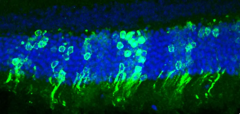
Professor Sowden’s previous research, showing a successful transplant of light-sensing cell into the retina of a blind mouse. This procedure improved the mouse’s vision and showed that this approach was a feasible way of treating inherited sight loss. Light-sensing rod cells are green, and the rest of the retina is blue (University College London).
What could this mean for our sight loss community?
This laboratory research is still at an early stage and will not lead directly to a treatment. However, if successful, this work could one day provide a way of restoring light-sensing cells in people with advanced vision loss. Unlike gene therapies, which are designed for specific genetic diagnoses, replacing light-sensing cells could help people with many different inherited retinal dystrophies. The researchers hope that the work will provide part of the evidence needed to support the first human clinical trials of their cell patches in the future.
Progress so far
One year into the project, Professor Sowden’s team has:
- Designed and produced patches strong enough to be handled during surgery and suitable for implantation beneath the retina.
- Worked with an ophthalmic technology company to develop a specialised surgical device that can place the patch beneath the retina more reliably than existing techniques.
- Successfully implanted the scaffold into the retinas of pigs, demonstrating that the procedure is feasible.
- Shown that implanted scaffolds can remain in place for at least a month without causing major structural damage to the retina.
- Grown large numbers of healthy light-sensing cells and purified them for transplantation.
Check back in early 2028 for the final outcomes of this project
Learn more about this research
Read more about Professor Jane Sowden’s research
Read about the gene faults that can cause inherited retinal dystrophies
Read about sight loss treatments that may work regardless of the genes involved
Read about Professor Sowden’s previous work to transplant light-sensing cells into mice retinas
Read about a clinical trial aiming to repair the retina with transplanted cells
Read more about why and how animals are used in scientific research
Read more about stem cells and how they’re used in retina research
- Lead researcher: Professor Rachael Pearson
- Location: King’s College London, England
- Type of funding: Project grant
- Total funding: £299,853
- Duration: October 2024 – April 2027
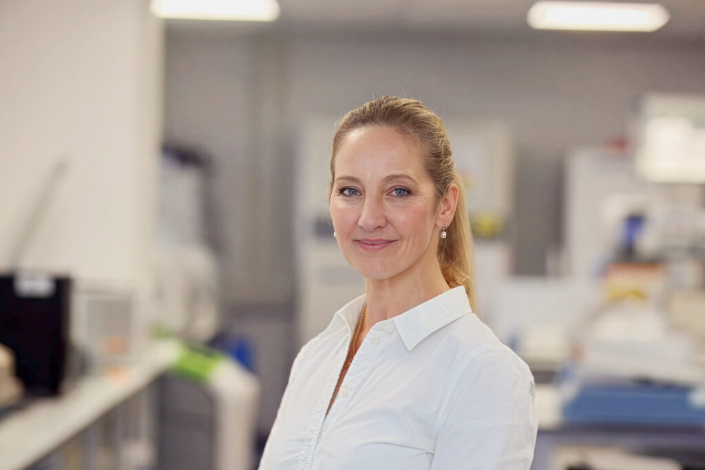
Professor Rachael Pearson, lead researcher (Retina UK).
The challenge
Scientists need to study living things to understand diseases, but it’s not safe or ethical to carry out every experiment on a person. That’s why experimental models like rats, fruit flies, and cells grown in petri dishes are vital for medical research. Every research model has strengths and limitations. More complex ones, like live animals, reproduce the effects of whole-body systems like hormones and immunity, but at a cost. Although there are strict regulations to make sure animal research is ethical and humane, some level of distress is inevitable. But simpler models that don’t involve animals still miss important aspects of human disease.
In 2011, researchers in Japan described the first method for growing mini retinas from stem cells in the lab. This important model has revolutionised our understanding of how the retina works and what can go wrong in retinal diseases. Since that remarkable breakthrough, scientists around the world have been growing more refined mini retinas. However, achieving the same level of organisation and functionality as the real thing is still out of reach.
The human retina is only as thick as a couple of sheets of paper, but it’s packed with over 120 cell types organised in ten layers. One of the biggest challenges with current mini retinas is maintaining a healthy population of ganglion cells, which process electrical signals and pass them through the optic nerve to the brain. Until we solve this problem, scientists can’t accurately study retinal diseases that affect this layer. And this bottleneck reduces our ability to predict how sight loss will progress and develop new treatments.
The vision
In London, Professor Rachael Pearson is finding new ways of growing mini retinas with working ganglion cells. Current models lack an optic nerve for the ganglion cells to latch onto, which might be why these cells don’t survive long term. Professor Pearson’s team have found that when they grow mini retinas on tiny plastic threads coated with a jelly of proteins and sugar, the ganglion cells stretch their nerve fibres along the threads. With this extra support, more of the ganglion cells survive for longer, suggesting this method could produce a model for studying them.
Now, Professor Pearson’s team are finetuning their mini retinas. By testing a variety of biodegradable thread materials and jelly coatings, they’re identifying the best blend to nurture ganglion cells. Once their new model is up and running, they plan to grow mini retinas with the gene fault that causes autosomal dominant optic atrophy. This inherited condition affects around 1 in 25,000 people in the UK and affects the optic nerve and ganglion cells of the retina. They want to show that their improved way of growing mini retinas works for studying this disease. This proof of concept should open up the possibility of using the new model to understand a much wider range of retinal conditions.
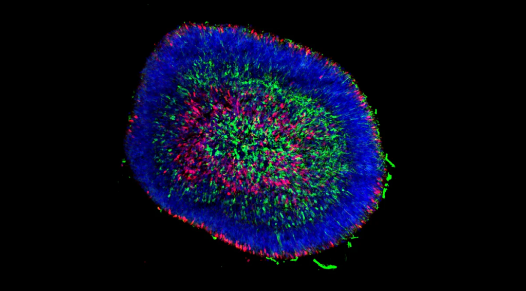
A microscopy image of a lab-grown mini retina about two-thirds of a millimetre across. The light-sensing cells have been stained red, the ganglion cells green, and all other cells blue (Kim Edwards, Stem Cell Reports).
What could this mean for our sight loss community?
This laboratory research is still at an early stage and will not lead directly to a treatment. However, if successful, this work could produce a new model for studying diseases that affect the retina. By allowing scientists to learn more about how inherited retinal dystrophies affect ganglion cells, the mini retinas Professor Pearson produces could help identify new treatment strategies targeting these cells. What’s more, if researchers test experimental drugs on her mini retinas, they may discover ways the drug works that a less advanced model would miss. And every improvement to mini retinas is another step towards reducing the number of animals used in research.
Progress so far
One year into the project, Professor Pearson’s team has:
- Designed and tested a new 3D-printed growth chamber which makes feeding their mini retinas and taking pictures of them easier.
- Identified a better thread material for their mini retinas grow nerve fibres along. They’re continuing to test other thread materials.
- Shown that coating their plastic threads in a certain protein encourages more nerve fibres to grow from the mini retinas.
- Started building their mini retina model of autosomal dominant optic atrophy.
Check back in mid-2027 for the final outcomes of this project.
Learn more about this research
Read more about why and how animals are used in scientific research
Read more about UK science that aims to reduce the number of animals needed for research
Read more about Professor Rachael Pearson’s research
Read more about mini organs (or organoids)
Read about the first mini retinas grown in 2011
Read about the 120 types of cells identified in the retina
- Lead researcher: Professor Omar Mahroo
- Location: University College London, England
- Type of funding: PhD studentship
- Total funding: £119,867
- Duration: October 2024 – October 2027

Professor Omar Mahroo, lead researcher (NIHR Moorfields BRC).
The challenge
Stargardt disease is the most common inherited macular dystrophy. It affects the macula, a small area near the centre of the retina that allows us to read, recognise faces, use digital screens, and see fine detail. As the condition progresses, an affected person’s central vision gradually deteriorates, making many everyday activities increasingly difficult.
Most research into Stargardt disease focuses on the loss of vision itself. However, many people with the condition also experience other symptoms that can be just as disruptive. Two of these symptoms are photopsia, the appearance of flashes or shapes of light that aren’t really there, and photophobia, an extreme sensitivity to bright light. These symptoms can interfere with daily life, yet they remain surprisingly poorly understood. Some people develop anxiety when flashes of light appear unexpectedly, while others struggle with bright environments or digital screens. Many also report frustration that these symptoms are not always recognised as part of Stargardt disease.
In a pilot study, more than 9 in 10 people with Stargardt disease reported experiencing photopsia, while over 3 in 10 reported photophobia. Despite being so common, no large study has investigated how often these symptoms occur, how they affect people, or what causes them.
The vision
In London, Professor Mahroo’s team wants to understand why photopsia and photophobia occur in Stargardt disease and whether they provide clues about how the condition develops over time.
The researchers will begin by surveying more than 100 people with Stargardt disease. They will ask participants about the flashes of light they see, how often they occur, what triggers them, and how much they affect everyday life. They will also investigate experiences of light sensitivity and how people cope with it.
Next, the team will compare these experiences with detailed measurements already collected during routine eye appointments. These include retina scans, functional tests, and measurements of vision. By combining these different sources of information, the researchers hope to identify patterns that link symptoms to specific stages of Stargardt disease.
The researchers will also carry out specialised, non-invasive vision tests to investigate how different retinal circuits are working. This could help them identify which cells in the retina are responsible for generating flashes of light and increased sensitivity to brightness.

A computer-generated illustration of photopsia, a symptom reported by many people with Stargardt disease. Photopsia can appear as flashes, sparkles, shimmering patterns, or glowing shapes that are seen despite no external light source being present. (Vision Center).
What could this mean for our sight loss community?
This project will not directly lead to a new treatment. However, it could provide something that many people with Stargardt disease have been asking for: recognition of their overlooked symptoms. Simply validating people’s experiences can have an important impact on their wellbeing.
By understanding when photopsia and photophobia occur, clinicians may be able to better explain what people can expect as their condition progresses and offer more tailored advice on managing these symptoms. The research may also reveal whether these symptoms are linked to particular patterns of retina damage or disease progression. If so, they could eventually help doctors predict how someone’s Stargardt disease may change over time or respond to treatment.
Although this study focuses on Stargardt disease, the findings could also benefit people with other inherited retinal dystrophies who experience similar symptoms.
Progress so far
Six months into the project, Professor Mahroo’s team has:
- Refined their patient survey to better capture experiences of how photopsia and photophobia affect everyday activities.
- Worked with the Stargardt community at the Stargardt’s Connected Annual Conference to gather feedback and encourage participation in the study.
- Expanded recruitment to include a more diverse and representative group of people with Stargardt disease.
- Found early evidence that photopsia and photophobia are common symptoms of Stargardt disease.
- Prepared access to clinical records from 69 genetically confirmed Stargardt disease patients.
Check back in early 2028 for the final outcomes of this project.
Learn more about this research
Read an interview with Hassina Zeriri, the PhD student working on this project
Read more about Professor Omar Mahroo’s research
- Lead researcher: Doctor Jörn Lakowski
- Location: University of Southampton, England
- Type of funding: Project grant
- Total funding: £243,747
- Duration: August 2024 – August 2027
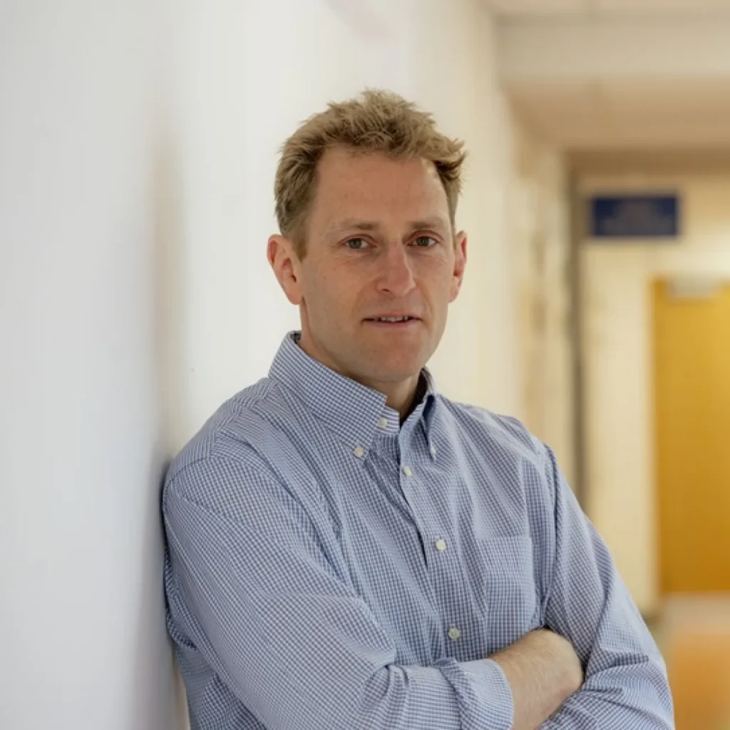
Doctor Jörn Lakowski, lead researcher (University of Southampton).
The challenge
Retinitis pigmentosa is the most common form of inherited sight loss, affecting 1 in 4,000 people in the UK. It usually begins with problems seeing in dim light and a gradual narrowing of the visual field, often described as “tunnel vision”. Over time, many people with retinitis pigmentosa also lose their central vision.
This pattern of sight loss is surprisingly puzzling. Most of the gene faults that cause retinitis pigmentosa directly affect only one type of light-sensing cell in the retina, called rods. Rods are responsible for night vision and most of our peripheral vision. Yet as the disease progresses, a second type of light-sensing cell also begins to die. These cells, called cones, provide our sharp central vision, colour vision, and ability to read, recognise faces, and see fine detail.
Scientists have known for many years that rod cells usually die before cone cells in retinitis pigmentosa. This has led to an intriguing theory: healthy rods may provide cones with a vital survival signal. When enough rods are lost, that support disappears, causing cones to slowly starve and die. Although this idea is widely accepted, much of the supporting evidence comes from animal studies. We still know surprisingly little about how cone starvation occurs in the human retina or how it might be prevented.
The vision
In Southampton, Doctor Lakowski’s team is using mini retinas grown from human stem cells to investigate why cone cells die in retinitis pigmentosa. These lab-grown models closely resemble the human retina and allow researchers to study disease processes that would otherwise be impossible to observe.
The researchers have developed a human model of cone starvation by switching off a gene called BSG1, which plays an important role in the survival pathway connecting rods and cones. They hope this model will reproduce some of the stresses experienced by cones during retinitis pigmentosa. As rod cells die, cone cells may lose access to critical support signals and nutrients. Dr Lakowski’s team wants to understand exactly what those missing factors are and how cone cells respond when they run short.
The researchers will compare healthy and starving cone cells to identify the biological pathways that help cones survive. They will then test more than 1,500 existing medicines to see whether any can keep starving cone cells alive for longer. Because most of these compounds are already approved for other conditions, promising candidates could potentially move towards clinical testing more quickly than entirely new drugs.
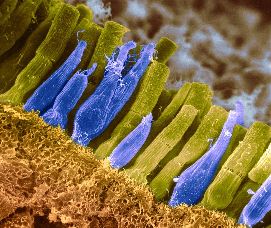
An image taken with an electron microscope showing rod and cone light-sensing cells. The picture has been falsely coloured on a computer so that rods appear yellow and cones blue. Beneath the cells is the retina’s external limiting membrane, which holds rods and cones in place. (Omikron, Pixels.com).
What could this mean for our sight loss community?
This laboratory research is still at an early stage and will not directly lead to a treatment. However, it could answer one of the most important unanswered questions in retinitis pigmentosa research: why do cone cells die after rod cells are lost?
If the team can identify the biological mechanisms that protect cones, researchers may be able to develop treatments that preserve central vision even after rod degeneration has begun. This could have a major impact on quality of life, helping people retain the vision needed for reading, recognising faces, and carrying out many everyday activities.
Importantly, cone loss occurs in most forms of retinitis pigmentosa regardless of which gene fault caused the condition. Therefore, any treatment that keeps cones alive has the potential to benefit a large proportion of the retinitis pigmentosa community, rather than only people with a specific genetic diagnosis.
Progress so far
Two years into the project, Professor Lakowski’s team has:
- Successfully grown mini retinas carrying the BSG1 gene fault and tracked their development for more than 250 days.
- Shown that removing BSG1 does not prevent the normal development and maintenance of cone cells under standard laboratory conditions, suggesting cones have previously unknown ways of surviving.
- Developed new methods for isolating and analysing light-sensing cells from mini retinas, laying the groundwork for detailed studies of cone starvation.
- Created the bioinformatics tools needed to compare gene activity in healthy and starving cone cells.
- Established two different drug-screening systems that will allow thousands of existing medicines to be tested for their ability to protect starving cone cells.
Check back in early 2028 for the final outcomes of this project.
Learn more about this research
Read an interview with Doctor Jörn Lakowski about this project
Read more about Doctor Lakowski’s research
Read our guide to retinitis pigmentosa
Read more about how rod cells feed cone cells to keep them alive
- Lead researcher: Professor Jacqueline van der Spuy
- Location: King’s College London, England
- Type of funding: Project grant
- Total funding: £299,635
- Duration: April 2024 – March 2027

Professor Jacqueline van der Spuy, lead researcher (University College London).
The challenge
Leber congenital amaurosis is a severe inherited retinal dystrophy that starts within the first few months of life and leads to blindness within a few years. The condition accounts for about 1 in every 30 cases of severe visual impairment in children. Scientists have identified at least 20 different types of Leber congenital amaurosis, each caused by a fault in a different gene. There’s currently only one treatment for this condition, called Luxturna. However, because Luxturna only works for people with a fault in a gene called RPE65, fewer than 100 people in the UK are likely to be eligible for it. That leaves the vast majority of people with Leber congenital amaurosis without any treatment.
Leber congenital amaurosis type 4 (LCA4) is caused by faults in a gene called AIPL1. This gene plays a critical role in phototransduction, the process by which the retina converts light into the electrical signals that allow people to see. The AIPL1 gene helps light-sensing cells control the levels of a signalling molecule called cyclic guanosine monophosphate, or cGMP. When AIPL1 is faulty, cGMP can build up to harmful levels.
Imagine driving across the country in a car that can only use first gear. At first, the car still moves, but soon it’s clear that something is wrong. The engine revs harder than it should, fuel is wasted, and every mile places extra strain on the vehicle. Eventually, parts begin to wear out and fail. Scientists think something similar happens in LCA4. Normally, the light-sensing cells of the retina constantly adjust to changing levels of light, using cGMP. But in LCA4, cGMP becomes stuck at unusually high levels, leaving the cells unable to respond to changing light conditions. Over time, the light-sensing cells become stressed, stop communicating properly with the rest of the retina, and eventually die.
Scientists still don’t fully understand how excess cGMP causes light-sensing cells to die. Filling this knowledge gap could reveal new ways of treating LCA4.
The vision
Using cells from real people with LCA4, Professor Jacqueline van der Spuy has created mini retinas with the same faults in the AIPL1 gene. By studying these lab-grown models, her London team is exploring the earliest things that go wrong because of excess cGMP. The researchers are investigating three possible ways that excess cGMP damages light-sensing cells: by disrupting calcium levels, interfering with communication between retinal cells, and triggering the cells to self-destruct.
Alongside this discovery research, the team are testing molecular mimics of cGMP as potential treatments for LCA4. These molecules, called cGMP analogues, have shown promise in other inherited retinal dystrophies. The molecules appear to block some of the harmful signals triggered by excess cGMP, helping retinal cells return to a healthier state. Professor van der Spuy is treating her mini retinas with cGMP analogues to see if they can correct the negative impact of excess cGMP.
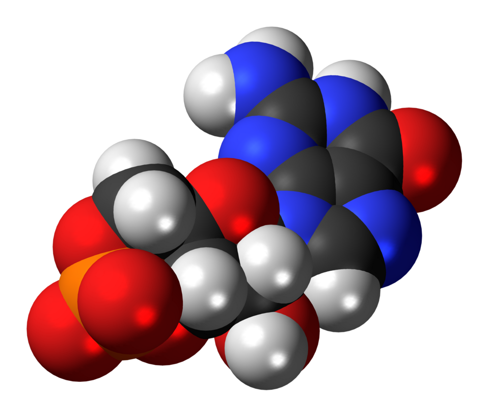
A three-dimensional representation of cGMP, the signalling molecule that gets stuck at abnormally high levels in people with LCA4. Atoms are shown as spheres: black for carbon, white for hydrogen, blue for nitrogen, red for oxygen, and orange for phosphorus. (Jynto, Wikipedia).
What could this mean for our sight loss community?
This laboratory research is still at an early stage and will not lead directly to a treatment. However, if successful, this work could identify new molecular mechanisms for researchers to target when designing treatments for LCA4. If the team sees promising results when treating mini retinas with cGMP analogues, this work could be an early step towards clinical trials for these molecules.
Abnormal cGMP levels in light-sensing cells aren’t just seen in LCA4, but in a wide variety of inherited retinal dystrophies. That means discoveries made in LCA4 could ultimately help researchers develop treatments for many different forms of inherited retinal dystrophy, including conditions caused by entirely different genes.
Progress so far
Two years into the project, Professor van der Spuy’s team has:
- Grown mini retinas from people with LCA4 and confirmed that they reproduce the harmful build-up of cGMP seen in the disease.
- Found evidence that communication between retinal cells becomes disrupted before the light-sensing cells begin to die.
- Developed new techniques to measure calcium levels in living mini retinas, helping them track how excess cGMP affects retinal function.
- Generated encouraging early evidence that a cGMP analogue reverses some of the harmful changes caused by excess cGMP.
- Started testing more cGMP analogues to identify the most promising candidates for future development.
Check back in mid-2027 for the final outcomes of this project.
Learn more about this research
Read more about Professor Jacqueline van der Spuy’s research
Read more about Leber congenital amaurosis
Read more about the estimated prevalence of Leber congenital amaurosis
Read more about cGMP and how controlling it can treat a range of diseases
Project start date: 2023
Prof Jacqueline van der Spuy, UCL Institute of Ophthalmology
Retina UK and the Macular Society are co-funding a PhD studentship to explore the use of prime editing to correct the most severe mutations causing Stargardt disease.
Prime editing is a cutting-edge technology in which a disease causing genetic change is precisely and permanently repaired in an individual’s genetic code, restoring the normal function of the gene. In this study, the researchers will investigate whether prime editing can efficiently correct one of the most common genetic changes causing Stargardt disease. By establishing methods for safely and precisely editing these particular faults, the project could contribute to the subsequent application of the technique across a huge range of conditions, potentially transforming future treatments for a wide proportion of those living with inherited sight loss.
This project is being co-funded by The Macular Society.
Learn more about completed research projects.